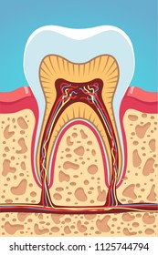
Bone Cross Section Diagram / Healthy tooth diagram isolated on white background vector.. Explaned distal and proximal epiphysis. A cross section of a human long bone. Ear external and internal anatomy cross section unlabeled stock illustration 9895a hr fotosearch / wh. There are trabeculae in spongy bone which gives its sponge like appearance. Bone marrow is the soft, highly vascular and flexible connective tissue within bone cavities which serve as the primary site of new blood cell production or hematopoiesis.
Cross section through middle metacarpal bones of vector. Two prominent grooves or sulci run along its length. Download 130+ royalty free bone cross section vector images. (micrograph provided by the regents of university of michigan. Cross section diagrams are used a lot by architects and engineers to show what a building or machine might look like before it's built.
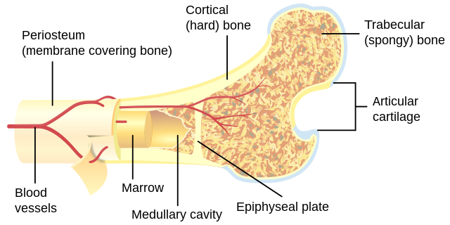
Cross Sectional Anatomy Kenhub from thumbor.kenhub.com Cross section diagrams are used a lot by architects and engineers to show what a building or machine might look like before it's built. Compact bone is the outer layer and the spongy bone forms the inner layer. For example, to read this diagram literally, since the cartilage can be seen inside the cutaway section of bone, it. Download 130+ royalty free bone cross section vector images. Find the perfect human bone cross section stock illustrations from getty images. Diagram with articular cartilage, marrow, medullary cavity and periosteum. Explaned distal and proximal epiphysis. Diagram with articular cartilage, marrow, spongy bone, medullary cavity, endosteum, diaphysis, and periosteum.
As shown in figure 2.
Vector illustration scheme of bone cross section. Healthy tooth diagram isolated on white background vector. Diagram with articular cartilage, marrow, spongy bone, medullary cavity, endosteum, diaphysis, and periosteum. Diagram with articular cartilage, marrow, medullary cavity and periosteum. For example, to read this diagram literally, since the cartilage can be seen inside the cutaway section of bone, it. Click to try our skeleton viewer. A cross section of a human long bone. Detailed and high textured 4k normal,disp,diffuse. Find the perfect human bone cross section stock illustrations from getty images. Vector illustration scheme of bone cross section. Ear external and internal anatomy cross section unlabeled stock illustration 9895a hr fotosearch / wh. Explaned distal and proximal epiphysis. Bone marrow is the soft, highly vascular and flexible connective tissue within bone cavities which serve as the primary site of new blood cell production or hematopoiesis.
Diagram with articular cartilage, marrow, spongy bone, medullary cavity, endosteum, diaphysis, and periosteum. Download 130+ royalty free bone cross section vector images. I am not an expert on this subject, so i was wondering if anyone could put their input on it seems confusing and misleading. Diagram with articular cartilage, marrow, spongy bone, medullary cavity, endosteum, diaphysis, and periosteum. Thin section of dinosaur bone.
Bone Cross Section High Res Stock Images Shutterstock from image.shutterstock.com Explaned distal and proximal epiphysis. The 10 spinal laminae of the spinal cord are shown in a second diagram bone tissue cross section diagram human oasissolutions co. For example, to read this diagram literally, since the cartilage can be seen inside the cutaway section of bone, it. Bone is found in the shafts of long bone and consists of various cylindrical units named as haversian system 47. Diagram with articular cartilage, marrow, spongy bone, medullary cavity, endosteum, diaphysis, and periosteum. Cross section through middle metacarpal bones of vector. The centroidal locations of common cross sections are well documented, so it is typically not necessary to calculate the location with the equations above. Labeled vertebra cross section of human body anatomy infographic diagram including all parts cord of grey and white matter spinal nerve vertebral body foramen and spinous process for medical.
Spinal cord spinal column anatomy information myvmc.
Compact bone is the outer layer and the spongy bone forms the inner layer. In a cross section of a bone we can see two types of bone tissue: From wikimedia commons, the free media repository. Spongy bone and compact bone. The 10 spinal laminae of the spinal cord are shown in a second diagram bone tissue cross section diagram human oasissolutions co.
Cross section through middle metacarpal bones of vector.
Labeled vertebra cross section of human body anatomy infographic diagram including all parts cord of grey and white matter spinal nerve vertebral body foramen and spinous process for medical. (micrograph provided by the regents of university of michigan. Thin section of dinosaur bone. Diagram with articular cartilage, marrow, medullary cavity and periosteum. Cross section diagrams are used a lot by architects and engineers to show what a building or machine might look like before it's built. Bone marrow is the soft, highly vascular and flexible connective tissue within bone cavities which serve as the primary site of new blood cell production or hematopoiesis. A cross section of a human long bone. Explaned distal and proximal epiphysis. There are trabeculae in spongy bone which gives its sponge like appearance. From wikimedia commons, the free media repository. Cross section of a bone diagram : Detailed and high textured 4k normal,disp,diffuse. Click to try our skeleton viewer.
Spongy bone and compact bone bone cross section. Jump to navigation jump to search.


0 Komentar